Pages

Subscribe:

Saturday 14 December 2013

MRI Scan Part-4

MRI Scan At A Glance

  • MRI scanning uses magnetism, radio waves, and a computer to produce images of body structures.
  • MRI scanning is painless and does not involve x-ray radiation.
  • Patients with heart pacemakers, metal implants, or metal chips or clips in or around the eyes cannot be scanned with MRI because of the effect of the magnet.
  • Claustrophobic sensation can occur with MRI scanning.

Friday 13 December 2013

MRI scan Part-3

How does a patient prepare for an MRI scan and how is it performed?

All metallic objects on the body are removed prior to obtaining an MRI scan. Occasionally, patients will be given a sedative medication to decrease anxiety and relax the patient during the MRI scan. MRI scanning requires that the patient lie still for best accuracy. Patients lie within a closed environment inside the magnetic machine. Relaxation is important during the procedure and patients are asked to breathe normally. Interaction with the MRI technologist is maintained throughout the test. There are loud, repetitive clicking noises which occur during the test as the scanning proceeds. Occasionally, patients require injections of liquid intravenously to enhance the images which are obtained. The MRI scanning time depends on the exact area of the body studied, but ranges from half an hour to an hour and a half.

How does a patient obtain the results of the MRI scan?

After the MRI scanning is completed, the computer generates visual images of the area of the body that was scanned. These images can be transferred to film (hard copy). A radiologist is a physician who is specially trained to interpret images of the body. The interpretation is transmitted in the form of a report to the practitioner who requested the MRI scan. The practitioner can then discuss the results with the patient and/or family.
Future
Scientists are developing newer MRI scanners that are smaller, portable devices. These new scanners apparently can be most useful in detecting infections and tumors of the soft tissues of the hands, feet, elbows, and knees. The application of these scanners to medical practice is now being tested.

Pictures of an MRI of the spine

This patient had a herniated disc between vertebrae L4 and L5. The resulting surgery was a discectomy
Picture of herniated disc between L4 and L5
Cross-section picture of herniated disc between L4 and L5

Thursday 12 December 2013

MRI scan Part-2

What are the risks of an MRI scan?


What are the risks of an MRI scan?


An MRI scan is a painless radiology technique that has the advantage of avoiding x-ray radiation exposure. There are no known side effects of an MRI scan. The benefits of an MRI scan relate to its precise accuracy in detecting structural abnormalities of the body.
Patients who have any metallic materials within the body must notify their physician prior to the examination or inform the MRI staff. Metallic chips, materials, surgical clips, or foreign material (artificial joints, metallic bone plates, or prosthetic devices, etc.) can significantly distort the images obtained by the MRI scanner. Patients who have heart pacemakers, metal implants, or metal chips or clips in or around the eyeballs cannot be scanned with an MRI because of the risk that the magnet may move the metal in these areas. Similarly, patients with artificial heart valves, metallic ear implants, bullet fragments, and chemotherapy or insulin pumps should not have MRI scanning.
During the MRI scan, patient lies in a closed area inside the magnetic tube. Some patients can experience a claustrophobic sensation during the procedure. Therefore, patients with any history of claustrophobia should relate this to the practitioner who is requesting the test, as well as the radiology staff. A mild sedative can be given prior to the MRI scan to help alleviate this feeling. It is customary that the MRI staff will be nearby during MRI scan. Furthermore, there is usually a means of communication with the staff (such as a buzzer held by the patient) which can be used for contact if the patient cannot tolerate the scan.

Wednesday 11 December 2013

MRI scan Part-1


What is an MRI scan?

An MRI (or magnetic resonance imaging) scan is a radiology technique that uses magnetism, radio waves, and a computer to produce images of body structures. The MRI scanner is a tube surrounded by a giant circular magnet. The patient is placed on a moveable bed that is inserted into the magnet. The magnet creates a strong magnetic field that aligns the protons of hydrogen atoms, which are then exposed to a beam of radio waves. This spins the various protons of the body, and they produce a faint signal that is detected by the receiver portion of the MRI scanner. The receiver information is processed by a computer, and an image is produced.
The image and resolution produced by MRI is quite detailed and can detect tiny changes of structures within the body. For some procedures, contrast agents, such as gadolinium, are used to increase the accuracy of the images.

When are MRI scans used?

An MRI scan can be used as an extremely accurate method of disease detection throughout the body. In the head, trauma to the brain can be seen as bleeding or swelling. Other abnormalities often found include brain aneurysms, stroke, tumors of the brain, as well as tumors or inflammation of the spine.
Neurosurgeons use an MRI scan not only in defining brain anatomy but in evaluating the integrity of the spinal cord after trauma. It is also used when considering problems associated with the vertebrae or intervertebral discs of the spine. An MRI scan can evaluate the structure of the heart and aorta, where it can detect aneurysms or tears.
It provides valuable information on glands and organs within the abdomen, and accurate information about the structure of the joints, soft tissues, and bones of the body. Often, surgery can be deferred or more accurately directed after knowing the results of an MRI scan.

Tuesday 10 December 2013

Ascites Part-4


What is the outlook (prognosis) for ascites?

The outlook on ascites primarily depends on its underlying cause and severity.
In general, the prognosis of malignant ascites is poor. Most cases have a mean survival time between 20 to 58 weeks, depending on the type of malignancy as shown by a group of investigators.
Ascites due to cirrhosis usually is a sign of advanced liver disease and it usually has a fair prognosis with a 3-year survival rate of about 75%.
Ascites due to heart failure has a fair prognosis as the patient may live years with appropriate treatments.

Monday 9 December 2013

Ascites part-3

What are the complications for ascites?

Some complications of ascites can be related to its size. The accumulation of fluid may cause breathing difficulties by compressing the diaphragm and formation of pleural effusion.
Infections are another serious complication of ascites. In patients with ascites related to portal hypertension, bacteria from the gut may spontaneously invade the peritoneal fluid (ascites) and cause an infection. This is called spontaneous bacterial peritonitis or SBP. Antibodies are rare in ascites and, therefore, the immune response in the ascitic fluid is very limited. The diagnosis of SBP is made by performing a paracentesis and analyzing the fluid for the number of white blood cells or evidence of bacterial growth.
Hepatorenal syndrome is a rare, but serious and potentially deadly (average survival rates range from 2 weeks to about 3 months) complication of ascites related to cirrhosis of the liver leading to progressive kidney failure. The exact mechanism of this syndrome is not well known, but it may result from shifts in fluids, impaired blood flow to the kidneys, overuse of diuretics, and administration of contrasts or drugs that may be harmful to the kidney.

Can ascites be prevented?


The prevention of ascites largely involves preventing the risk factors of the underlying conditions leading to ascites.
In patients with known advanced liver disease and cirrhosis of any cause, avoidance of alcohol intake can markedly reduce the risk of forming ascites. Nonsteroidal antiinflammatory drugs (ibuprofen [Advil, Motrin, etc.]) should also be limited in patients with cirrhosis as they may diminish the blood flow to the kidneys, thus, limiting the salt and water excretion. Complying with dietary salt restrictions is also another simple preventive measure to reduce ascites.

Sunday 8 December 2013

Ascites Part-2

What is the treatment for ascites?

The treatment of ascites largely depends on the underlying cause. For example, peritoneal carcinomatosis or malignant ascites may be treated by surgical resection of the cancer and chemotherapy, while management of ascites related to heart failure is directed toward treating heart failure with medical management and dietary restrictions.
Because cirrhosis of the liver is the main cause of ascites, it will be the main focus of this section.

Diet

Managing ascites in patients with cirrhosis typically involves limiting dietary sodium intake and the use of diuretics (water pills). Restricting dietary sodium (salt) intake to less than 2 grams per day is very practical, successful, and widely recommended for patients with ascites. In majority of cases, this approach needs to be combined with the use of diuretics as salt restriction alone is generally not an effective way to treat ascites. Consultation with a nutrition expert in regards to daily salt restriction can be very helpful for patients with ascites.

Medication

Diuretics increase water and salt excretion from the kidneys. The recommended diuretic regimen in the setting of liver related ascites is a combination of spironolactone (Aldactone) and furosemide (Lasix). Single daily dose of 100 milligrams of spironolactone and 40 milligrams of furosemide is the usual recommended initial dosage. This can be gradually increased to obtain appropriate response to the maximum dosage of 400 milligrams of spironolactone and 160 milligrams of furosemide, as long as the patient can tolerate the dose increase without any side effects. Taking these medications together in the morning is typically advised to prevent frequent urination during the night.

Therapeutic paracentesis

For patients who do not respond well to or cannot tolerate the above regimen, frequent therapeutic paracentesis (a needle carefully is placed into the abdominal area, under sterile conditions) can be performed to remove large amounts of fluids. A few liters (up to 4 to 5 liters) of fluid can be removed safely by this procedure each time. For patients with malignant ascites, this procedure may also be more effective than diuretic use.

Surgery

For more refractory cases, surgical procedures may be necessary to control the ascites. Transjugular intrahepatic portosystemic shunts (TIPS) is procedure done through the internal jugular vein (the main vein in the neck) under local anesthesia by an interventional radiologist. A shunt is placed between the portal venous system and the systemic venous system (veins returning blood back to the heart), thereby reducing the portal pressure. This procedure is reserved for patients who have minimal response to aggressive medical treatment. It has been shown to reduce ascites and either limit or eliminate the use of diuretics in a majority of cases performed. However, it is associated with significant complications such as hepatic encephalopathy (confusion) and even death.
Mor

Saturday 7 December 2013

Ascites Part-1

Ascites Facts

  • Ascites refers to abnormal accumulation fluid in the abdominal (peritoneal) cavity.
  • The most common cause of ascites is cirrhosis of the liver.
  • Treatment of ascites depends on its underlying cause.

What is ascites?

Ascites is the accumulation of fluid (usually serous fluid which is a pale yellow and clear fluid) in the abdominal (peritoneal) cavity. The abdominal cavity is located below the chest cavity, separated from it by the diaphragm. Ascitic fluid can have many sources such as liver disease, cancers, congestive heart failure, or kidney failure.

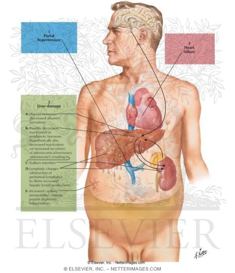
What causes ascites?

The most common cause of ascites is advanced liver disease or cirrhosis. Approximately 85% of the ascites cases are thought to be due to cirrhosis. Although the exact mechanism of ascites development is not completely understood, most theories suggest portal hypertension (increased pressure in the liver blood flow) as the main contributor. The basic principle is similar to the formation of edema elsewhere in the body due to an imbalance of pressure between inside the circulation (high pressure system) and outside, in this case, the abdominal cavity (low pressure space). The increase in portal blood pressure and decrease in albumin (a protein that is carried in the blood) may be responsible in forming the pressure gradient and resulting in abdominal ascites.
Other factors that may contribute to ascites are salt and water retention. The circulating blood volume may be perceived low by the sensors in the kidneys as the formation of ascites may deplete some volume from the blood. This signals the kidneys to reabsorb more salt and water to compensate for the volume loss.
Some other causes of ascites related to increased pressure gradient are congestive heart failure and advanced kidney failure due to generalized retention of fluid in the body.
In rare cases, increased pressure in the portal system can be caused by internal or external obstruction of the portal vessel, resulting in portal hypertension without cirrhosis. Examples of this can be a mass (or tumor) pressing on the portal vessels from inside the abdominal cavity or blood clot formation in the portal vessel obstructing the normal flow and increasing the pressure in the vessel (for example, the Budd-Chiari syndrome).
Acites can also manifest as a result of cancers, called malignant ascites. These types of ascites are typically manifestations of advanced cancers of the organs in the abdominal cavity, such as, colon cancer, pancreatic cancer, stomach cancer, breast cancer, lymphoma, lung cancer, or ovarian cancer.
Pancreatic ascites can be seen in people with chronic (long standing) pancreatitis or inflammation of the pancreas. The most common cause of chronic pancreatitis is prolonged alcohol abuse. Pancreatic ascites can also be caused by acute pancreatitis as well as trauma to the pancreas.

Friday 6 December 2013

Ascites Cont.

When should I call my doctor about ascites?

People with ascites should be routinely followed by their primary physician and any specialists that may be involved in their care. Gastroenterologists (specialists in gastrointestinal diseases) and hepatologist (liver specialists) commonly see patients with ascites due to liver disease. Other specialists can also care for patients with ascites based on the possible cause and the underlying condition. The specialists usually ask the patient to first contact their primary physician if ascites increase. If ascites is causing symptoms of shortness of breath, abdominal discomfort ,or inability to do normal daily tasks such as walking, the patient's primary doctor should be notified.

How is ascites diagnosed?

The diagnosis of ascites is based on physical examination in conjunction with a detailed medical history to ascertain the possible underlying causes since ascites is often considered a nonspecific symptom for other diseases. If ascites fluid is greater than 500ml, it can be demonstrated on physical examination by bulging flanks and fluid waves performed by the doctor examining the abdomen. Smaller amounts of fluid may be detected by an ultrasound of the abdomen. Occasionally, ascites is found incidentally by an ultrasound or a CT scan done for evaluating other conditions.
Diagnosis of underlying condition causing ascites is the most important part of understanding the reason for a person to develop ascites. The medical history may provide clues to the underlying cause and typically includes questions about previous diagnosis of liver disease, viral hepatitis infection and its risk factors, alcohol abuse, family history of liver disease, heart failure, cancer history, and medication history.
Blood work can play an essential role in evaluating the cause of ascites. A complete metabolic panel can detect patterns of liver injury, functional status of the liver and kidney, and electrolyte levels. A complete blood count is also useful by providing clues to underlying conditions. Coagulation (clotting) panel abnormalities (prothrombin time) may be abnormal because of liver dysfunction and inadequate production of clotting proteins.
Sometimes the possible underlying causes of ascites may not be determined based on the history, examination, and review of laboratory data and imaging studies. Analysis of the fluid may be necessary in order to obtain further diagnostic data. This procedure is called paracentesis, and it is performed by trained physicians. It involves sterilizing an area on the abdomen and, with the guidance of ultrasound, inserting a needle into the abdominal cavity and withdrawing fluid for further analysis.
For di

Thursday 5 December 2013

Ascites

What are the types of ascites?

Traditionally, ascites is divided into 2 types; transudative or exudative. This classification is based on the amount of protein found in the fluid.
A more useful system has been developed based on the amount of albumin in the ascitic fluid compared to the serum albumin (albumin measured in the blood). This is called the Serum Ascites Albumin Gradient or SAAG.
  • Ascites related to portal hypertension (cirrhosis, congestive heart failure, Budd-Chiari) is generally greater than 1.1.
  • Ascites caused by other reasons (malignant, pancreatitis) is lower than 1.1.

What are the risk factors for ascites?

The most common cause of ascites is cirrhosis of the liver. Many of the risk factors for developing ascites and cirrhosis are similar. The most common risk factors include hepatitis B, hepatitis C, and long standing alcohol abuse. Other potential risk factors are related to the other underlying conditions, such as congestive heart failure, malignancy, and kidney disease.

What are the symptoms of ascites?


There may be no symptoms associated with ascites especially if it is mild (usually less than about 100 – 400 ml in adults). As more fluid accumulates, increased abdominal girth and size are commonly seen. Abdominal pain, discomfort, and bloating are also frequently seen as ascites becomes larger. Shortness of breath can also happen with large ascites due to increased pressure on the diaphragm and the migration of the fluid across the diaphragm causing pleural effusions (fluid around the lungs). A cosmetically disfiguring large belly, due to ascites, is also a common concern of some patients.

Wednesday 4 December 2013

Anemia Part-8


Take the Blood Disorders Quiz

What are the complications of anemia?

As mentioned earlier, hemoglobin has the important role of delivering oxygen to all parts of the body for consumption and carries back carbon dioxide back to the lung to exhale it out of the body. If the hemoglobin level is too low, this process may be impaired, resulting in low levels of oxygen in the body (hypoxia).

What is the outlook (prognosis) for anemia?

Anemia generally has a very good prognosis and it may be curable in many instances. The overall prognosis depends on the underlying cause of anemia, its severity, and the overall health of the patient.

Tuesday 3 December 2013

Anemia Part-7

Anemia Part-7

Take the Blood Disorders Quiz

How is blood collected for a CBC?

Blood is collected by venipuncture (using a needle to draw blood from a vein) in a lab, hospital, or physician's office. Typically, blood is collected in a special sterile tube from an arm vein. The tube has some preservatives to prevent clotting of the blood. Results may be available in an hour or longer depending on the setting.
In some instances, a quick in office test called hemoglobin rapid test may be performed using a few drops of blood from a finger prick. The advantage of this quick test is that results may be obtained in a few minutes and only a few drops of blood may be required.

What is the red blood cell (RBC) count?

The red blood cells (RBCs or erythrocytes) are the most common type of cells in the blood. We each have millions and millions of these little disc-shaped cells. The RBC count is done to determine if the number of red blood cells is low (anemia) or high (polycythemia).
In an RBC count, the number and size of the RBCs are determined. This is usually reported as number of RBCs per a specified volume, typically in millions of RBCs in microliters (one one-thousandth of an ml) of whole blood. The shape of the RBCs is also evaluated under a microscope. All of this information, the number, size and shape of the RBCs, is useful in the diagnosis of anemia. Further, the specific type of anemia may be determined by this information.

What is hemoglobin?

Hemoglobin is a red pigment that imparts the familiar red color to red blood cells and to blood. Functionally, hemoglobin is the key chemical compound that combines with oxygen from the lungs and carries the oxygen from the lungs to cells throughout the body. Oxygen is essential for all cells in the body to produce energy.
The blood also transports carbon dioxide, which is the waste product of this energy production process, back to the lungs from which it is exhaled into the air. The transport of the carbon dioxide back to the lung is also achieved by hemoglobin. The carbon dioxide bound to hemoglobin is unloaded in the lungs in exchange for oxygen to be transported to the tissues of the body.

Monday 2 December 2013

Anemia Part-6

Take the Blood Disorders Quiz

What does a low hemoglobin level mean?

Low hemoglobin is called anemia. When there is a low hemoglobin level, there is often a low red blood cell count and a low hematocrit, too. Reference ranges are slightly different from one source to another, but typically hemoglobin of less than 13.5 gram/100 ml is abnormal in men and less than 12.0 gram/100 ml in women.

What is the hematocrit?

The hematocrit is specifically a measure of how much of the blood is made of red cells. The hematocrit is a very convenient way to determine whether the red blood cell count is too high, too low, or normal. The hematocrit is a measure of the proportion of blood that is composed of the red blood cells.

How is hematocrit determined?

The red blood cells in the sample of blood are packed down by spinning the tube in a centrifuge under prescribed conditions. The proportion of the tube that consists of red blood cells is then measured. Let's say that it is 45%. The hematocrit is 45%.

How is anemia treated?

The treatment of the anemia varies greatly. First, the underlying cause of the anemia should be identified and corrected. For example, anemia as a result of blood loss from a stomach ulcer should begin with medications to heal the ulcer. Likewise, surgery is often necessary to remove a colon cancer that is causing chronic blood loss and anemia.
Sometimes iron supplements will also be needed to correct iron deficiency. In severe anemia, blood transfusions may be necessary. Vitamin B12 injections will be necessary for patients suffering from pernicious anemia or other causes of B12 deficiency.
In certain patients with bone marrow disease (or bone marrow damage from chemotherapy) or patients with kidney failure, epoetin alfa (Procrit, Epogen) may be used to stimulate bone marrow red blood cell production.
If a medication is thought to be the culprit, then it should be discontinued under the direction of the prescribing doctor.

Anemia Part-5

Take the Blood Disorders Quiz

How is anemia diagnosed?

Anemia is usually detected, or at least confirmed, by a complete blood cell (CBC) count. A CBC test may be ordered by a physician as a part of routine general checkup and screening or based on clinical signs and symptoms that may suggest anemia or other blood abnormalities.

What is a complete blood cell (CBC) count?

Traditionally, CBC analysis was performed by a physician or a laboratory technician by viewing a glass slide prepared from a blood sample under a microscope. Today, much of this work is often automated and done by machines. Six component measurements make up a CBC test:
  1. Red blood cell (RBC) count
  2. Hematocrit
  3. Hemoglobin
  4. White blood cell (WBC) count
  5. Differential blood count (the "diff")
  6. Platelet count
Only the first three of these tests -- the red blood cell (RBC) count, the hematocrit, and the hemoglobin -- are relevant to the diagnosis of anemia.
Additionally, mean corpuscular volume (MCV) is also often reported in a CBC, which basically measures the average volume of red blood cells in a blood sample. This is important in distinguishing the causes of anemia. Units of MCV are reported in femtoliters, a fraction of one millionth of a liter.
Other useful clues to causes of anemia that are reported in a CBC are the size, shape, and color of red blood cells.
Picture of Red Blood Cells

Sunday 1 December 2013

Anemia Part-4

Take the Blood Disorders Quiz

Yes, anemia may be genetic. Hereditary disorders can shorten the life span of the red blood cell and lead to anemia (for example, sickle cell anemia). Hereditary disorders can also cause anemia by impairing the production of hemoglobin (for example, alpha thalassemia and beta thalassemia).
Depending on the degree of the genetic abnormality, hereditary anemias may cause mild, moderate, or severe anemia. In fact, some may be too severe to be compatible with life and may result in death of the fetus (unborn infant). On the other hand, some of these anemias are so mild that they are not noticeable and are incidentally revealed during a routine blood work.

What are the symptoms of anemia?

Some patients with anemia have no symptoms. Others with anemia may feel:
  • Tired
  • Fatigue easily
  • Appear pale
  • Develop palpitations (feeling of heart racing)
  • Become short of breath
Additional symptoms may include:
  • Hair loss
  • Malaise (general sense of feeling unwell)
  • Worsening of heart problems
It is worth noting that if anemia is longstanding (chronic anemia), the body may adjust to low oxygen levels and the individual may not feel different unless the anemia becomes severe. On the other hand, if the anemia occurs rapidly (acute anemia), the patient may experience significant symptoms relatively quickly.